What is the image quality of Animal Digital Dental DR after multiple uses?
Leave a message
As a supplier of Animal Digital Dental DR, I've received numerous inquiries about the image quality of our product after multiple uses. In this blog, I'll delve into this topic, drawing on our extensive experience and in - depth knowledge of the technology.
Understanding Animal Digital Dental DR
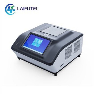
Before discussing the long - term image quality, it's essential to understand what Animal Digital Dental DR is. This advanced imaging technology is specifically designed for veterinary dental applications. It offers a digital alternative to traditional film - based dental X - rays, providing veterinarians with clear, detailed images of an animal's teeth and surrounding structures.
Unlike traditional X - ray systems, Animal Digital Dental DR uses a digital detector to capture X - ray images. These detectors are highly sensitive and can convert X - ray photons into electrical signals, which are then processed into digital images. This digital format allows for easy storage, sharing, and manipulation of the images, enhancing the diagnostic capabilities of veterinarians.
Factors Affecting Image Quality in Initial Use
When a new Animal Digital Dental DR system is first used, several factors contribute to its image quality. Firstly, the detector's sensitivity plays a crucial role. High - quality detectors can capture more X - ray photons, resulting in clearer and more detailed images. The spatial resolution of the detector also matters. A higher spatial resolution means that smaller details in the dental structures can be accurately represented in the image.
Another important factor is the X - ray source. A well - calibrated X - ray source can provide a consistent and appropriate dose of radiation. This ensures that the images are neither over - exposed nor under - exposed, maintaining optimal contrast and sharpness. Additionally, the software used to process the captured images can enhance the image quality by adjusting parameters such as brightness, contrast, and color.
Image Quality After Multiple Uses
Over time, with multiple uses, the question arises: does the image quality of Animal Digital Dental DR deteriorate? In general, if the system is properly maintained, the impact on image quality is minimal.


One of the main concerns is the wear and tear of the detector. The detector is the heart of the Animal Digital Dental DR system, and repeated exposure to X - rays can potentially cause damage. However, modern detectors are designed to be highly durable. They are made from materials that can withstand a large number of X - ray exposures without significant degradation. For example, some detectors use amorphous silicon or cesium iodide technology, which have excellent stability and long - term performance.
The X - ray source also needs to be considered. With continuous use, the X - ray tube may experience some changes in its output. However, regular calibration can ensure that the X - ray source maintains a consistent radiation output. Most Animal Digital Dental DR systems are equipped with self - calibration features or can be easily calibrated by trained technicians. This calibration process compensates for any minor changes in the X - ray tube's performance, ensuring that the image quality remains stable.
The software used in the system also evolves over time. Software updates are often released by the manufacturers to improve the image processing algorithms. These updates can enhance the image quality by reducing noise, improving contrast, and providing more accurate diagnostic information. As a result, even after multiple uses, the image quality can be maintained or even improved with the help of software enhancements.
Comparing with Other Veterinary Imaging Technologies
To better understand the long - term image quality of Animal Digital Dental DR, it's useful to compare it with other veterinary imaging technologies. Animal Imaging Diagnostic CT provides three - dimensional images of the animal's body, including the dental area. While CT can offer more detailed anatomical information, it is also more expensive and requires more complex equipment. In terms of long - term image quality, CT systems also face challenges such as detector degradation and the need for regular calibration.
Handheld Veterinary Dental X Ray is a more portable option. However, its image quality may be more limited compared to Animal Digital Dental DR. Handheld systems often have lower spatial resolution and may be more prone to image artifacts due to their size and portability. After multiple uses, the image quality of handheld systems may also be affected by factors such as battery performance and detector wear.
Maintaining Image Quality
To ensure that the Animal Digital Dental DR system maintains its image quality after multiple uses, proper maintenance is essential. This includes regular cleaning of the detector and the X - ray source to prevent dust and debris from affecting the image quality. The system should also be stored in a suitable environment, away from extreme temperatures and humidity.
Regular servicing by trained technicians is also recommended. During servicing, the system can be thoroughly inspected, and any potential issues can be addressed before they affect the image quality. Additionally, following the manufacturer's guidelines for use and maintenance is crucial. This includes using the correct exposure settings, avoiding unnecessary X - ray exposures, and performing regular software updates.
Real - World Examples
In our experience as a supplier, we have seen many veterinary clinics using our Animal Digital Dental DR systems for years without significant degradation in image quality. For example, a small animal clinic in a rural area has been using our system for over five years. They perform multiple dental X - rays every day, and the images they obtain are still of high quality. The clinic's veterinarian attributes this to the system's durability and the regular maintenance they perform.
Another example is a large - scale veterinary hospital. They have multiple Animal Digital Dental DR systems in use. By following a strict maintenance schedule and regularly updating the software, they have been able to maintain consistent image quality across all their systems. This has allowed them to provide accurate diagnoses and high - quality dental care to their patients.
Conclusion
In conclusion, the image quality of Animal Digital Dental DR after multiple uses remains high if the system is properly maintained. The detector's durability, the X - ray source's calibration, and the software's continuous improvement all contribute to the long - term stability of the image quality.
If you are a veterinarian or a veterinary clinic owner considering purchasing an Animal Digital Dental DR system, you can be confident in its long - term performance. Our company is committed to providing high - quality products and excellent after - sales service. If you have any questions or are interested in discussing a purchase, please feel free to reach out to us. We look forward to the opportunity to work with you and help you enhance your veterinary dental imaging capabilities.
References
- Manufacturer's documentation of Animal Digital Dental DR systems
- Research papers on veterinary imaging technology and detector durability
- Case studies from veterinary clinics using Animal Digital Dental DR systems