What preparation is required for an animal before an imaging diagnostic CT scan?
Leave a message
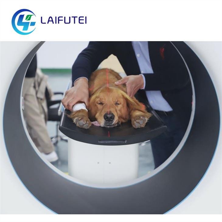
Preparing an animal for an imaging diagnostic CT scan is a meticulous process that requires careful consideration of various factors to ensure accurate results and the well - being of the animal. As a supplier of Animal Imaging Diagnostic CT, I understand the importance of proper preparation. In this blog, I will outline the key steps and preparations necessary for an animal before undergoing a CT scan.
1. Initial Consultation and Medical History
The first step in preparing an animal for a CT scan is an in - depth consultation with the veterinarian. The vet will review the animal's medical history, including any pre - existing conditions, previous surgeries, allergies, and current medications. This information is crucial as it can affect the scan process and the interpretation of the results. For example, if an animal has a history of kidney problems, the use of contrast agents during the CT scan may need to be carefully evaluated, as these agents can put additional stress on the kidneys.
During the consultation, the vet will also conduct a physical examination of the animal. This helps to identify any visible signs of illness or injury that may be relevant to the scan. The vet will assess the animal's vital signs, such as heart rate, respiratory rate, and temperature, to ensure that the animal is in a stable condition to undergo the CT scan.
2. Fasting
Fasting is a common preparation step for animals before a CT scan, especially when a contrast agent is to be used. Fasting helps to reduce the amount of gas and food in the digestive tract, which can interfere with the clarity of the images. For most animals, a fasting period of 8 - 12 hours is recommended. However, the exact fasting time may vary depending on the species, age, and health condition of the animal.
For example, puppies and kittens may not be able to tolerate long fasting periods, so the fasting time may need to be adjusted accordingly. It is important to follow the veterinarian's instructions regarding fasting to ensure the best possible scan results.
3. Sedation or Anesthesia
CT scans require the animal to remain still for an extended period. To achieve this, sedation or anesthesia is often necessary, especially for small animals or those that are likely to be anxious or uncooperative. The type of sedation or anesthesia used will depend on the animal's species, age, health condition, and the nature of the scan.
Veterinarians will carefully select the appropriate sedative or anesthetic agent to minimize the risk of complications. Before administering sedation or anesthesia, the vet will perform a pre - anesthetic evaluation, which may include blood tests and other diagnostic procedures to assess the animal's organ function and overall health. This helps to ensure that the animal can safely tolerate the sedation or anesthesia.
4. Shaving and Cleaning
If the area of interest for the CT scan is covered with fur, the veterinarian may need to shave the fur to improve the quality of the images. Shaving the fur reduces artifacts caused by the scattering of X - rays by the hair. The area to be shaved will be determined based on the location of the suspected problem.
After shaving, the area will be cleaned to remove any dirt, debris, or oils from the skin. This helps to ensure that the contrast agent, if used, can be properly absorbed and that the skin surface is clean for better image quality.
5. Contrast Agent Administration
In some cases, a contrast agent may be used during the CT scan to enhance the visibility of certain tissues and blood vessels. Contrast agents work by increasing the density of the tissues they are in, making them more distinguishable on the CT images.
There are different types of contrast agents available, including intravenous (IV) contrast agents and oral contrast agents. IV contrast agents are injected into the animal's bloodstream, while oral contrast agents are given by mouth. The choice of contrast agent and the method of administration will depend on the specific diagnostic needs.
Before administering the contrast agent, the veterinarian will perform a thorough assessment of the animal's health to ensure that the animal can tolerate the contrast agent. The vet will also monitor the animal closely during and after the administration of the contrast agent for any signs of adverse reactions, such as allergic reactions or kidney problems.
6. Collar and Leash Removal
Before the CT scan, any collars, leashes, or other metal objects on the animal should be removed. Metal objects can cause artifacts on the CT images, which can interfere with the accurate interpretation of the results. It is important to ensure that the animal is free of any metal items that could affect the scan.
7. Communication with the Owner
Throughout the preparation process, clear communication with the animal owner is essential. The veterinarian should explain the purpose of the CT scan, the preparation steps, and the potential risks and benefits to the owner. The owner should be informed about the fasting requirements, the need for sedation or anesthesia, and what to expect during and after the scan.
The owner should also be given instructions on how to care for the animal after the scan. This may include information on post - anesthetic recovery, such as monitoring the animal's behavior, appetite, and urination. The owner should be encouraged to ask any questions they may have to ensure that they are fully informed and comfortable with the process.
8. Equipment Preparation
As a supplier of Animal Imaging Diagnostic CT, we understand the importance of having well - maintained and calibrated equipment. Before each scan, the CT scanner should be checked to ensure that it is functioning properly. The scanner should be calibrated regularly to ensure accurate and consistent image quality.
The appropriate scan protocols should be selected based on the animal's species, size, and the area of interest. Different animals may require different scan parameters, such as tube voltage, tube current, and slice thickness, to obtain the best possible images.
In addition to the CT scanner, other equipment such as the Image Scanner CR may also be used in conjunction with the CT scan for further image processing and analysis. The Image Scanner CR can help to enhance the quality of the images and provide additional diagnostic information.

9. Staff Training
The staff involved in the CT scan process, including veterinarians, technicians, and nurses, should be well - trained in animal handling, anesthesia administration, and CT scan operation. Proper training ensures that the animal is treated with care and that the scan is performed safely and accurately.
Staff should be familiar with the different types of CT scanners and their operating procedures. They should also be trained in recognizing and managing any potential complications that may arise during the scan, such as adverse reactions to sedation or anesthesia.
10. Quality Assurance
Quality assurance is an important part of the CT scan process. After the scan, the images should be reviewed by a qualified veterinarian or radiologist to ensure that they are of sufficient quality for diagnosis. The images should be free of artifacts and should clearly show the area of interest.
If the images are not of adequate quality, the scan may need to be repeated. Quality assurance also involves maintaining accurate records of the scan, including the scan parameters, the use of contrast agents, and the results of the image review.
Contact for Procurement
If you are interested in purchasing high - quality Animal Imaging Diagnostic CT equipment for your veterinary practice, we are here to assist you. Our products are designed to provide accurate and reliable diagnostic images, and our team of experts can offer comprehensive support and training. Please feel free to reach out to us to start a procurement discussion. We also offer related products such as the Image Scanner CR and Animal Digital Dental DR to meet your diverse diagnostic needs.
References
- Thrall, D. E., Robertson, J. D., & Biery, D. N. (2012). Textbook of Veterinary Diagnostic Radiology. Elsevier Health Sciences.
- Flecknell, P. A. (2015). Laboratory Animal Anaesthesia. Academic Press.
- Proceeding of the American College of Veterinary Radiology annual meetings.